FS/2/2/2/1/11 – Selected papers from veterinary case notes relating to Ovarian Tumours
This material is Crown copyright, and contains public sector information licensed under the Open Government License v.3.0.
Click here for full transcript of Case Notes
[FS/2/2/2/1/11]
[[1]]
Ovarian Tumour
Notes on Case
[Annotated ‘Bay Mare S 30 6 yrs old 2 years service’]
1 ½ gallon fluid in abdomen
Pelvic Flexure of colon lying against Diaphragm anterior to Tumour.
Circumference 55 inches – 44 inches short circumference
48 inches
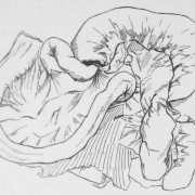
[Pencil sketch with measurements]
[Annotated ‘The greatest diameter & smallest of the tumour should be given that conveying more to the mind of its size than the weight &c]
Colon at pelvic flexure & caecum at its head covered with innumerable blood clots – seen also on the Peritoneum generally, but more especially on the floor of abdomen.
Weight 70 lbs [Annotated ‘How long since first ill’]
Thorax contained a quantity of fluid which could not be weighed
Anterior to left lung a large tumour, evidently Bronchial Lymphatic Glands attached to Pericardium, there being some gelatinous material between the Glands & Pericardm. Weight of tumour 6lbs. Sac around tumour pretty tough, but consistence of latter of thick custard pudding, blood coloured. Adherence between sac & substance of tumour. Left Lung healthy – Right Lung slightly emphysematous at its inferior border. Heart – Pale ext[erna]ly, substance firm. Blood patch in anr.vent. furrow. Semilunar valves thickened, mitral valves healthy. Aortic valves healthy.
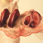
Ovarian Tumour Weight 70 lbs. Appearance on section like putrid cheese both in consistence & color[sic], one part, breaking down had occurred, the cysts containing thick coffee colored[sic] material – the amount being about 14 oz. The sac itself was very firm, in some places ½ in. in thickness. Externally principally yellowish, especially towards floor of abdomen: in other parts it had suffered from the general peritonitis.
Diaphragm much thickened, covered on its anterior by scrofulous deposits on its posterior, peritonitic blood clots: Lumbar glands much enlarged, size of Bombay mango: on section, structure broken down – like parotid gland somewhat.
Right Kidney weight 1 lbs. Section healthy. Left Kidney healthy
Pre Renal capsule enlarged
Spleen – 1 ¼ lbs – Lymphatics along G.S Omentum very much enlarged, swelling out & some softness. Spleen capsule blood stained in portions from peritonitis.
Liver 15 lbs about. Covered with peritonitic lymph. Portal vessels surrounded by scrofulous deposits – no deposits in its substance.
Stomach – Deposits around greater curvature. Otherwise healthy. Large intestine collapsed containing dark green offensive mucous. Muc[ous] mem[brane] of colon much congested. Caecum healthy
[[2]]
[Sketch of ovarian tumour in abdomen]
Mesentery covered with Peritonitic lymph. Glands not much enlarged.
[Transcription by Claudia Watts, KCL History, April 2019]





Leave a Reply
Want to join the discussion?Feel free to contribute!