FS/2/2/2/2/2 – Selected papers from veterinary case notes relating to Joint Diseases
This material is Crown copyright, and contains public sector information licensed under the Open Government License v.3.0.
Click here for full transcript of Case Notes for Horse 23
[FS/2/2/2/2/2]
[[1]]
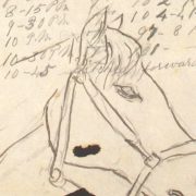
5/1st R.A
Case 30, No 23
[Annotated ‘Lameness’]
1883
Augt 13th
Sprain to sesamoid[sic] sheath of off hind limb. Extreme lameness and pain. Foment and give Aloes ℨV – Place in slings
[August] 15
Physic acting – The animal is in great pain – high heeled shoe
[August] 24
Placed in slings – suffering acutely – hot fomentations
Sept 1st
Still in great pain – There must be some very severe injury to this sheath – continue treatment – Removed from slings being badly chafed
Sept 20th
Not a big better and an abcess[sic] has formed at the fetlock, discharging a little thin pus. Fomentations is the only thing which gives relief the animal is losing flesh and considering the length of time he has been ill it is a wonder he has not died of irritative fever
Oct 10th
The animal do not make the least improvement he is constantly holding the limb in the air. The sheath is tense, thickened, hot, painful and another abcess[sic] has formed on the outside of the fetlock, also discharging a little thin pus & synovia.
[October] 23rd
I cannot make out the case, it is wonderful that an animal should for so many weeks suffer such acute pain with no sign of abatement, I believe the sesamoid sheath is ruptured and I will try the counter irritating effect of a blister – Put back in slings and blister the part. The suffering is telling
[[2]]
upon the poor animal who is daily loosing flesh though on full feed, if he were not a young horse I would certainly apply for his destruction.
26th
The blister has acted as yet with no good effect
28th
Still as painful as ever, the limb is held up all day
Nov 20th
I think there is a little improvement, he bears more weight on it and is looking better.
The brush[?] is enormously distended, tense & painful – He has been now in slings a month
[November] 27
Continues to improve
Decr 24th
An abcess[sic] which does not communicate with the brush has formed and burst on the external Surface of the bursa or is about the size of a Rupee. Dress all with dry bran. Stands much better. I am not likely to remain in charge much longer or I would certainly fine the joint as soon as this abcess[sic] has healed.
[Handwriting changes]
1884, 28 Jan
When I saw this animal to day he was going sound. The sheath fetlock is much enlarged but the [iron] should put him right. He is quite fat & plump now.
May 1
saw him again to day, not lame but has small abscess forming occasionally[?] around the pastern. Crow has fired him again & he still remains sound.
[Transcription by Claudia Watts, KCL History, April 2019]
Click here for full transcript of Case Notes for Horse 111
[FS/2/2/2/2/2]
[[1]]
Extract from Record
Case 133, No-111
1st Novb 1882
Wound incised off fore above Fetlock caused while jumping, the perforatus is nearly divided the synovial sac opened dress with Carbolized Bran & syringe every morning for an hour with Carbolized Water.
6th [November]
An abscess has formed in the heel owing to suppuration within the bursa. Continue treatment.
20th [November]
Wound healing well. Continue treatment.
Dec 1st
Nearly healed, a deal of thickening remains & horse
Looseing[sic] condition. Full feed, omit carbolic syringing & the carbolized bran
Jan 1st 1883
Wound healed the thickening is as great as ever would blister but consider it useless, what makes matters worse is the irritable condition of the patient who won’t go in slings & won’t lie quietly but continues hopping round the stall with a high heeled shoe on remove the latter & bring up for casting.
Extract from Register.
No 111 – Chestnut gelding; 4 years old; Australian.
Q/1st B[riga]de R.A
[Transcription by Claudia Watts, KCL History, April 2019]
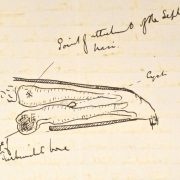
Click here for full transcript of Painting of Perforatus and Perforans of Horse 111
[FS/2/2/2/2/2]
[[1]]
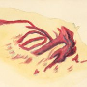
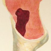
[Image – Left] Perforatus [Image – Right] Perforans
Injury to tendon caused by an over reach. Horse belonged to 2/1 RA no 111
The perforatus was thickened which extended around the joint. The perforatus, cut through, infected. The perforans infected, small portion of example is parts showing that it had become adherent.
[Transcription by Claudia Watts, KCL History, April 2019]