Posts

28 – Letter to Fred Bullock from Frederick Smith, 28 Mar 1913

Terms of Use
The copyright of this material belongs to the Royal College of Veterinary Surgeons. It is available for reuse under a Creative Commons, Attribution, Non-commercial license.

77 – Letter to Fred Bullock from Frederick Smith, 17 Jul 1913

Terms of Use
The copyright of this material belongs to the Royal College of Veterinary Surgeons. It is available for reuse under a Creative Commons, Attribution, Non-commercial license.

9 – Letter to Fred Bullock from Frederick Smith, 13 Feb 1914

Terms of Use
The copyright of this material belongs to the Royal College of Veterinary Surgeons. It is available for reuse under a Creative Commons, Attribution, Non-commercial license.

18 – Letter to Fred Bullock from Frederick Smith, 3 Mar 1914

Terms of Use
The copyright of this material belongs to the Royal College of Veterinary Surgeons. It is available for reuse under a Creative Commons, Attribution, Non-commercial license.

28 – Letter to Fred Bullock from Frederick Smith, 28 May 1922

Terms of Use
The copyright of this material belongs to the Royal College of Veterinary Surgeons. It is available for reuse under a Creative Commons, Attribution, Non-commercial license.

45 – Letter to Fred Bullock from Frederick Smith, 8 Nov 1922

Terms of Use
The copyright of this material belongs to the Royal College of Veterinary Surgeons. It is available for reuse under a Creative Commons, Attribution, Non-commercial license.

A model hoof

During the recent reordering of the RCVS headquarters a 6 inch square box was found containing a cardboard model and a folded piece of paper.  Further investigation revealed it was something rather exciting – the pasteboard model of a horse’s hoof which accompanies Bracy Clark’s two page pamphlet A new exposition of the horses’ hoof.

Bracy Clark (1771-1860), the son of a Quaker, was born in Chipping Norton, Oxfordshire.  According to Frederick Smith, in The early history of veterinary literature and its British Development Vol III, Clark  left school at 14 and was apprenticed to  a surgeon for seven years.    With the founding of the London Veterinary School in 1791 his thoughts turned to studying veterinary, rather than human, medicine and he enrolled at the London school some time during 1792, gaining his Diploma in July 1794.

A new exposition of the horses hoof

Signature and date

After a period touring the continent he opened a practice in Giltspur Street, London which mainly dealt with brewery horses.  Whilst in practice Clark developed an interest in shoeing, establishing a number of forges throughout the country. He was later joined at the practice by his nephew Charles Clark and appears to have retired from active work in 1828.

In retirement Clark, now settled at Taunton Street near Regents Park, devoted his energies to publishing and re-editing his own works and experimenting with shoeing.

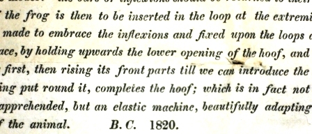
The pamphlet found in the box A new exposition of the horses’ hoof  is dated 1820 and signed BC.   It starts:

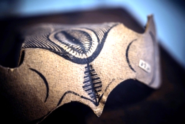
“It would be difficult by words or description alone to convey a correct notion of the framing and construction of the Horses’ hoof: I have therefore invented a pasteboard model, which exhibits its nature and properties very familiarly.

In order to understand it, it will be necessary to take it to pieces a few times, and put it together again, when the simplicity and manner of its construction will be strongly and clearly impressed on the mind”

Model to accompany A new exposition of the horses hoof

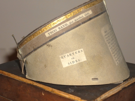
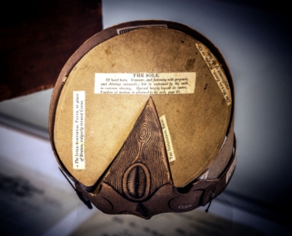
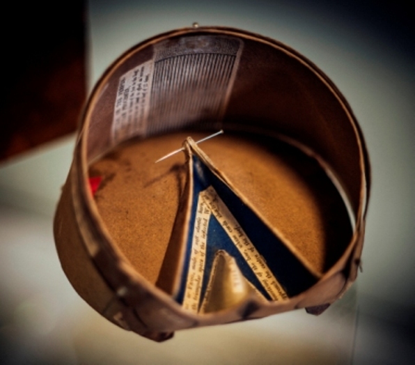
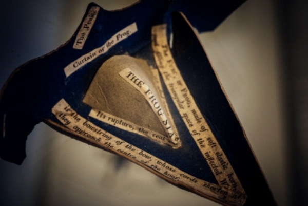
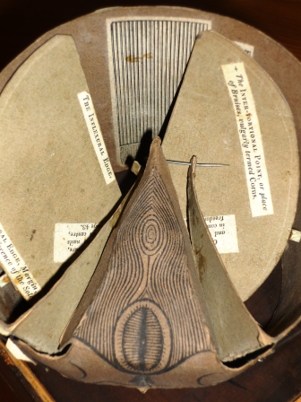
Clark explains how to dismantle the model by withdrawing the pins (which can be seen in the photographs) and how to learn more of the structure of the hoof by studying how the labelled flaps connect and move etc.
Model to accompany A new exposition of the horses hoof

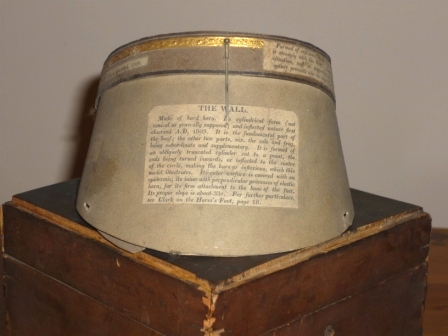
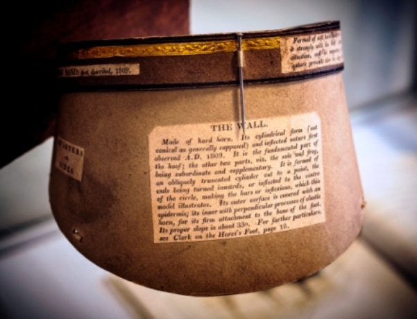
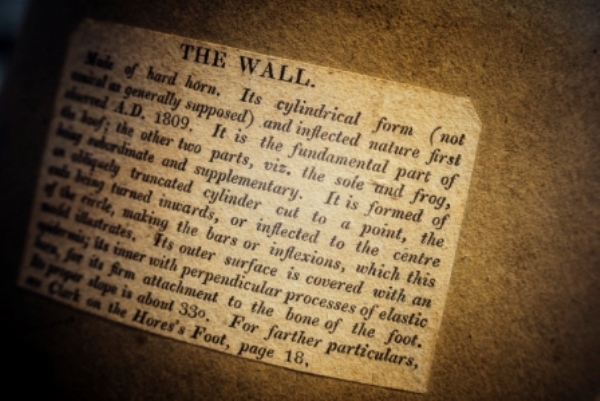
For example writing of the wall of the hoof which has been exposed by the removal of the frog and the sole from the model:

“by reversing the situation of the Bars or Inflexions,…we now discover that they are simply a continuation of the wall, obliquely growing narrower…We can now discern, that the wall and bars are one continued piece…of an obliquely cut cylinder…and the great simplicity and power of such an arrangement must call forth our exulted admiration…”

Model to accompany A new exposition of the horses hoof

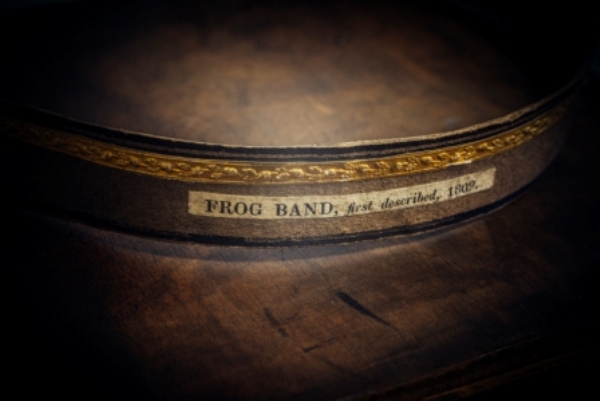
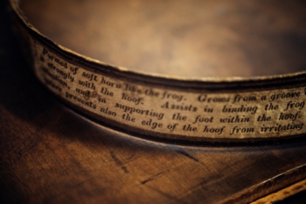
The final paragraph of the pamphlet which explains how to put the model back together concludes:

“Now the Frog-band…completes the hoof: which is in fact not merely a rude covering of horn, as been apprehended, but an elastic machine, beautifully adapting itself to all degrees of exertion, or repose of the animal.”

Considering the model may be nearly 200 years old it is in very good condition – though as yet I have not been brave enough to take it apart.  This is due to the fact that it had its own lockable box – which in itself gives some indication of its value at the time the pamphlet was written.

At the moment we do not know how it comes to be in our possession but we do know that Bracy Clark  gave a complete set of his works to the RCVS library around the time he became a Vice President of the College in 1857.  So the ‘hoof’ could be Clark’s very own model, we will let you know what we find out!

Strickland Freeman’s observations on the horses foot

Our historical collection has over two hundred books on farriery and horse shoeing many of which contain anatomical drawings of the foot of the horse.  One such book is Strickland Freeman’s

Observations on the mechanism of the horse’s foot its natural spring explained, and a mode of shoeing recommended, by which the foot is defended from external injury, with the least impediment to its spring

which was published in 1796.

Strickland  Freeman was a Buckinghamshire landowner and sporting gentleman  who wrote a number of works on horsemanship and farriery.   Observations is his most important anatomical work: focusing on horse shoeing practices and methods, the work consists of  109 pages of text and 16 coloured plates with outlines.

I have found two reviews of the book from the period.  One in The Critical Review is less than complimentary.   Speaking of the illustrations it says:

“[they] do not illustrate the complete anatomy of the foot:  they are imperfect, from a total neglect of the nerves and absorbent vessels of those parts, two points in the structure, which have been found to be inseparably connected with many diseases of the feet of horses”

The reviewer also takes issue with the cost of the book given its alleged shortcomings:

 “When a voluminous and expensive work is laid before the public…we are led to expect something approaching toward a complete account of that subject… To the anatomist he affords no information, to the gentleman, we fear, he conveys little knowledge which can be applicable in practice… We would advise further adventurers in scientific pursuits with which they are not fully conversant, to be more backward in taxing the world with expensive books”.

A more positive review appeared in The Monthly Review which describes Observations as a ‘most splendid work’ and says:

“it is evidently the result of attentive observation…that will afford useful hints to those who are practically concerned in the subject”.

Writing in 1929 Frederick Smith in Volume 2 of his Early history of veterinary literature says:

“[Freeman] knew nothing of the anatomy of the horse’s foot.  The text is consistently weak, though largely atoned for by…[the] beautifully coloured plates”.

I am not qualified to comment on the accuracy of either the text or illustrations, but can’t help but agree that the illustrations, which are by G Kirtland who was a leading anatomical artist at that time, are beautiful.  Four of them are shown below, what do you think?

Plate 3. Front view of the bones of the fore foot of a horse in their relative situation

Plate 3. Front view of the bones of the fore foot of a horse in their relative situation

Plate 4. Back view of the bones of the fore foot in their relative situation

Plate 4. Back view of the bones of the fore foot in their relative situation

Plate 8. View of the posterior surface of the foot to shew the arteries and veins

Plate 8. View of the posterior surface of the foot to shew the arteries and veins

Plate 9. Side view of the foot to shew the arteries and veins

Plate 9. Side view of the foot to shew the arteries and veins

Bracy Clark’s hoof – an update

I am delighted to say that thanks to the generosity of members of the Central Veterinary Society we have been able to have some conservation work carried out on Bracy Clark’s pasteboard model hoof.  At the same time the surface dirt was removed which means that you can now read the labels much more clearly as you can see in the photographs below.

We are also very grateful to colleagues at the Royal Veterinary College who have kindly produced this 3D model – we hope you enjoy exploring the hoof as much as we do.

Bracy Clark hoof - the wall

21863175559_c24de7a26b_o

Bracy Clark hoof _underside

Bracy Clark hoof - the wall

Bracy Clark hoof - curtain

Bracy Clark hoof

Bracy Clark hoof - frog band

Bracy Clark hoof