Oxford Street’s pioneering veterinary surgeon

London, like most towns or cities, has lots of plaques commemorating people or events attached to its buildings.  Most of these plaques are above eye level and often go unnoticed by people passing by.

Site of plaque commemorating William Moorcroft

Site of plaque commemorating William Moorcroft

One such plaque is sited on the left-hand side of Oxford Street, when walking down from Marble Arch, on the block just before Portman Street.  As you can see from the photo above – it is quite hard to spot as it is small, high up and surrounded by garish shop displays.

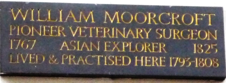
A close up of the plaque, which was erected by the Veterinary History Society in 1982, reveals that it commemorates the fact that William Moorcroft, veterinary surgeon, lived and practiced on the site from 1793-1808.

Plaque on the site of William Moorcroft's practice

Plaque on the site of William Moorcroft’s practice

William Moorcroft 1767-1825 was born in Ormskirk, Lancashire the grandson of a wealthy landowner and farmer.  In the early 1780s he was apprenticed to a surgeon in Liverpool but during an outbreak of an unknown cattle disease he was recruited to help treat the stricken animals.  Moorcroft impressed the local landowners so much that they offered to underwrite his education if he gave up his plans to study surgery and went instead to the veterinary school in Lyon, France.

This he did, arriving in France in 1789, graduating a year or so later as the first Englishman to qualify as a veterinary surgeon.

Returning to London he set up in practice at 224 Oxford Street (the street has since been renumbered). In 1794 Moorcroft was made joint Professor, with Edward Coleman, of the London Veterinary College. However the arrangement only lasted a few weeks before he resigned seemingly because of the difficulties of combining academic life with a thriving business.  The Oxford Street practice continued to grow, with Moorcroft recruiting John Field, firstly as his assistant and then later as his partner, until it became one of the most lucrative in London.

As well as his work in the practice Moorcroft also purchased breeding stock for the East India Company and in 1803 was appointed manager of their stud in Essex.  Then in 1808 he was appointed as Superintendent of the East India Company’s stud in Bengal at a salary of £3,000 a year (the size of the salary offered to Moorcroft gives some indication of just how successful the practice must have been).

Thus began the part of his life for which Moorcroft is perhaps better known.  As well as improving the control of disease in the stud in Bengal he established a number of subsidiary studs and introduced a co-operative breeding programme with the local population.   However the breeding programme still did not produce the type of horse that was needed for cavalry and artillery purposes and Moorcroft began to look further afield.

In 1811 he travelled some 1500 miles across North India looking for new breeding stock. Then in May 1812 he set out on a second journey, with Captain Hyder Hearsey, this time looking for both horses and as a side issue the Tibetan shawl goat.  (Moorcroft saw the goat as offering an opportunity for increased trade between the company and the locals).

Their journey took them through Kumaon and the Niti Pass into Tibet, finally ending at Lake Manasarovar, and resulted in the purchase of Kashmir goats, some of which eventually ended up in Scotland, but little in the way of horses.  Perhaps not surprisingly Moorcroft’s employers were not impressed – his primary purpose had been to look for horses and he came back with goats! – and they curtailed his travels for a number of years.

In 1819 Moorcroft  embarked on what was to be his final – 6 year long – journey.  Visiting Ladakh (1820-1822), Kasmir and the Punjab (1822-1823), Afghanistan (1824-1824), Turkestan (1824-1825), finally reaching Bokhara, in modern Uzbekistan, on 25th February 1825.   He died from fever on the return journey on 27 August 1825.

William Moorcroft, – pioneering veterinary surgeon and Asian explorer – quite a story which the plaque only hints at.  Want to find out more? Then check out some of the books listed below.

Further reading

Alder, Garry (1985) Beyond Bokhara : The Life Of William Moorcroft, Asian Explorer And Pioneer Veterinary Surgeon, 1767-1825  London: Century Publishing

Irwin, John (1973) The Kashmir shawl   London : HMSO

Travels in the Himalayan Provinces of Hindustan and the Punjab, in Ladakh and Kashnair, in Peshawur, Kabul, Kunduz and Bokhara, from 1819 to 1825: by William Moorcroft and George Trebeck:  prepared from the press from original journals and correspondence by H. H. Wilson.  London: 1845 (reprinted  New Delhi : Sagar Publications 1971)  

The artistic Mr Mayhew

I have recently had the opportunity to look more closely at the almost 400 watercolours by Edward Mayhew that we acquired in 1990 and which are now available to view on our Vet History site

Edward Mayhew (1808-1868) was the brother of Henry Mayhew, founding editor of Punch. It is thought he spent his early years in the theatre before enrolling at the London Veterinary College in 1843 when he was in his 30s. He qualified as a member of the RCVS on 6 February 1845 and became a member of RCVS Council just over a year later.

Reports of the meetings in the three years Mayhew spent on Council show him to be a man who was not afraid to speak his mind, especially where the Professors at both the London and Edinburgh veterinary schools were concerned. His rather short obituary in The Veterinarian (November 1868 p810) says very little about him except “He was well known as the author of several veterinary works.”

It is for two of these veterinary works, Illustrated horse doctor  and Illustrated horse management, that the watercolours were prepared and most of them have counterparts in the published books.

The drawings, which were converted into woodcuts for publication, reveal a man of no small artistic talent.

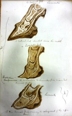
The collection includes illustrations:  of diseases including the stages of laminitis

The stages of laminitis

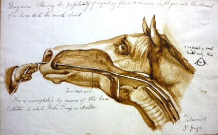
methods of treatment – inserting a tube into the stomach of a horse

Inserting a tube into the stomach of a horse

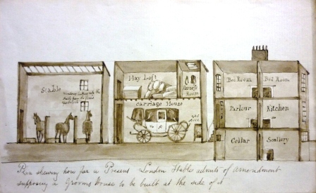
and show how horses, and their keepers, should be housed

Plan showing how a stable can have a grooms house attached alongside

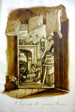
They also reveal something of the social conditions at the time – including this ‘Peep into the Grooms Home’

A Peep into the Grooms Home

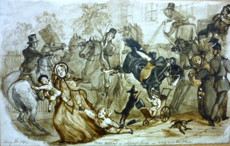
My favourites are the ones which show Mayhew’s  sense of humour – for example this drawing titled ‘Never mount a strange horse in a crowded place.’ I particularly like the ‘flying’ dog on the bottom right!

Never mount a strange horse in a crowded place

 Update (25/8/15)

Whilst researching for my lastest blog post on Mayhew I found an interesting account of the Mayhew family in George Hodder’s  Memories of my time: including personal reminiscences of eminent men  (London: Tinsley Brothers, 1870)

The section on Edward Mayhew (pp58-62)  gives information on his health when producing these illustrations  and mentions that his brother Julius, an architect, helped with the illustrations and designs for the stables.

Links to digitised images

Images used in Illustrated Horse Doctor

Images used in Illustrated Horse Management

Images that do not appear in published works

One of the professions youngest and brightest ornaments

Recently there has been another of those coincidences that those of us who work in libraries love.  Two quite separate enquiries which end up having something – or in this case someone – in common.

The first enquiry was from a researcher who wanted to look at The quarterly journal of veterinary science in India and army animal management and the second from someone who was researching James McCall, founder of Glasgow Veterinary College.

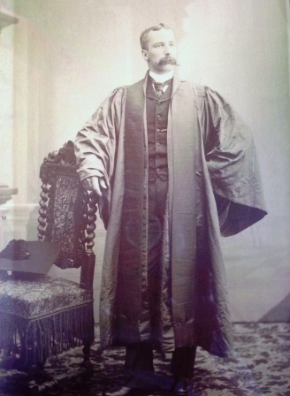
John Henry Steel

John Henry Steel

The connection? John Henry Steel – who was the co-founder of the journal and who had a medal named after him which was awarded to McCall in 1899.

As is usual when this happens curiosity got the better of me and I had to find out more..

John Henry Steel FRCVS (1855-1891) followed his father into the veterinary profession, graduating from the London Veterinary College in 1875.  After a brief spell in the army he took up a post as Demonstrator of Anatomy at the London College where he remained for five years, before resigning when the professorship, which he had been promised, was abolished.
He re-entered the army and, in 1882, went to serve in India, which was to be the scene of his most notable achievements. Upon arrival, he “was immediately impressed by the utter want, outside the Army, of anything approaching Veterinary Science” and set about rectifying the situation.

One of his first moves was to establish, in 1883, The quarterly journal of veterinary science in India and army animal management.  This journal, which he co-founded with Frederick Smith, allowed vets to share information and record  progress in treatments etc.

Secondly, in 1886, he established a veterinary college in Bombay so that the population could be “educated in veterinary matters”.

Sadly the severe mental and physical strain of running the journal and the College took its toll and he was taken ill, returning to England in 1888.  Against the advice of doctors, he went back to India after a few months and involved himself fully in the life of Bombay, taking up the reins at the College again and becoming a Fellow of the University and a JP.

His health did not improve and, seemingly aware of his imminent death, he wrote an editorial for the journal, dated October 1890, titled ‘Cui Bono’ (to whose benefit?).  It starts:

“To every conscientious worker there arrive times of introspection when the questions arise to him what has been the outcome of my efforts?”

Steel then proceeds to assess his life’s work and in particular to question the usefulness of the Journal and whether it was worth the “at times laborious work”.

He feels

“it has succeeded in enlarging the mind of the public and profession on matters veterinary…[and has] enabled men working on the same subjects… to co-ordinate their work and results”

and if at all possible it must continue

“Considering how things were before the Quarterly, considering the work our Journal has been enabled to do…we have decided to continue its production”

Announcement of closure of journal

Announcement of closure of journal

Sadly this was not to be and  Steel wrote an announcement  stating that owing to severe illness he was having to leave India and that the Journal will no longer be continued after December 1890.  The publisher then adds to the end of the announcement, which was distributed with the last issue, “J H Steel, Esq …died at Bombay on Thursday the 8th”.

He was  just short of his 36th birthday.

The three page obituary in the Veterinary Record (31 January 1891), from which the title of this post is taken, and the fact that the RCVS instituted a medal in his honour testifies to the high regard in which he was held by the profession.

The College which he founded, the Journal and his books on diseases of the ox, dog, elephant, sheep, and camel and on equine relapsing fever remain as his lasting legacy.

Edward Snape’s muscular preparation

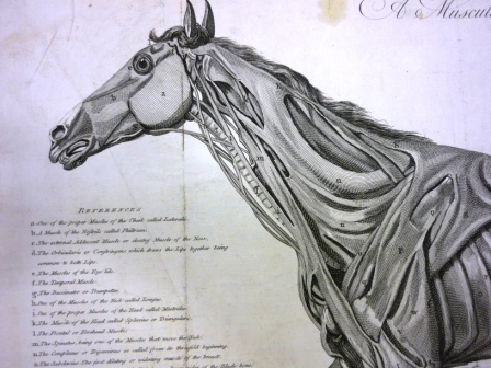
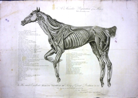
The library is currently having a makeover which has meant emptying shelves and cupboards.  One thing that has come out of its cupboard is a print titled ‘A muscular preparation of a horse with references.’

Snape A Muscular preparation of a horse

Edward Snape – A Muscular preparation of a horse

The inscription reads:

“To His most Excellent MAJESTY GEORGE III. King of Great Britain &c &c. This Plate is most humbly inscribed by His Majesty’s most faithfull (sic) Subject and Servant Edwd Snape Farrier to their Majesty’s & the 2nd Troop of the Horse Guards.
Published 15th April 1778 and sold by E. Snape in Berkeley Square”

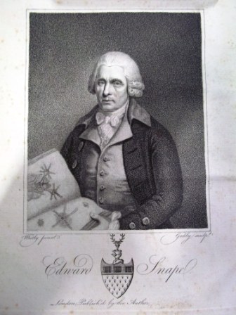
Edward Snape 1728-18?? was a London based farrier who claimed to be a descendant of Andrew Snape of The Anatomy of an horse fame.

Edward Snape

Edward Snape

Little is known of him but he seems to have had a genuine desire to see his profession progress.  In 1766 he proposed the establishment of a “hippiatric [horse] infirmary” as a school for the “instruction of pupils in the profession.”   The school eventually came into existence in Knightsbridge in 1778.

There is a short account of the school in an ‘advertisement’ (which reads like an obituary though Snape was still alive and presumably the publisher knew this!) in the 2nd, 1805,  edition of the Practical treatise on farriery (1st edition 1791) though it doesn’t actually say it opened –  just that it was built.  The ‘advertisement’ also makes the following claim “whatever benefit the Country now derives from the establishment of the Veterinary College, originated in him.”

Alongside the Practical treatise the print is the only other thing that Snape is known to have published.  My research suggests that  Snape’s print is based on an earlier  one by Jeremiah Bridges.  Looking at reproductions of the two prints side by side in the Veterinary History article the illustrations are remarkably similar though Snape has added labels (his ‘with references’).  If it is true that Snape has copied an existing illustration then he would share a trait in common with his supposed ancestor Andrew Snape who used Carlo Ruini’s Anatomia del Cavallo as the basis for his work (for more information on Ruini’s work and Andrew Snape)

Close up of head

Close up of head

It would appear that the print I ‘found’ is not the first ‘edition’ as it has some minor changes to the inscription when compared to the earliest known copies.  Luckily we also have a copy of the earlier printing (of which Wood says there are only 4 known copies)

One interesting thing about the early version of the print is that  whilst it bears the date 1778 all the known copies are on paper bearing an 1808 watermark. I doubt we will ever know why that is the case.  It could it be that it was prepared for use at the hippiatric infirmary but not actually printed until the later date.  Or was it produced in 1808 but ‘back dated’ to give some credence to the claim that the benefits of the Veterinary College (now the Royal Veterinary College, founded 1791) originated in Snape?

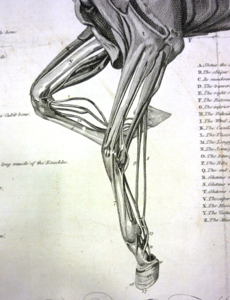
Close up fore legs

Close up – fore legs

Once the library is back in order I will give the two prints a proper outing and put them on display – do come and see them.

Update: See comments for further information on Snape

Bibliography

Smith, Frederick (1924) The Early history of veterinary literature and its British development Vol II.  London: Bailliere, Tindall & Cox

Wood, John GP (2004) A tale of two prints: Jeremiah Bridges and Edward Snape Veterinary History Vol 12 no 2 pp 173-183

Open wide

In a paper read before members of the Veterinary Medical Association (probably in 1849) Thomas Gowing said

“I have …experienced great difficulties in many required operations on the teeth [of the horse] from the want of proper dental instruments.”

His solution?  To invent a set of instruments which “I flatter myself, will be found useful… [and] I venture to recommend them to you with confidence.”

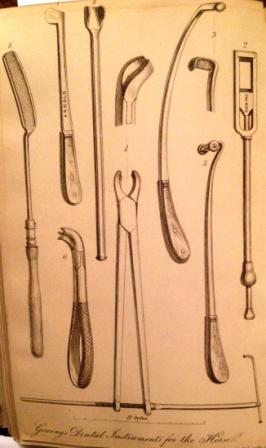
The set included: large and small forceps; a sliding chisel; a guarded chisel; lateral repellers; posterior repellers; a gum lancet, and a tooth rasp.

Thomas Gowing's dental instruments

Thomas Gowing’s dental instruments

His paper was later published as An essay, descriptive and pathological, on the diseases incidental to the horse; containing remarks upon operative dental surgery and the instruments required The 13 page essay contains an illustration of this dental set (above), some discussion of the possible causes of tooth disease in horses and explanations of how to use the instruments.

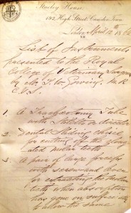
List of dental instruments donated by Thomas Gowing

List of dental instruments donated by Thomas Gowing

Thomas William Gowing 1810?-1888 was a London practitioner who qualified from the London Veterinary College in 1847.   He served on RCVS Council for over twenty years, including periods as Vice President and was one of the Primary Fellows  elected in 1877.

He gave a slightly modified set of these instruments to the RCVS Museum in 1866 – as recorded in a note which is bound within a book of pamphlets in our collection alongside his paper to the VMA.

I have yet to find any other illustrations of mid 19th century dental instruments in our historical collections but images of  teeth appear frequently, usually as part of discussions on how to age a horse.

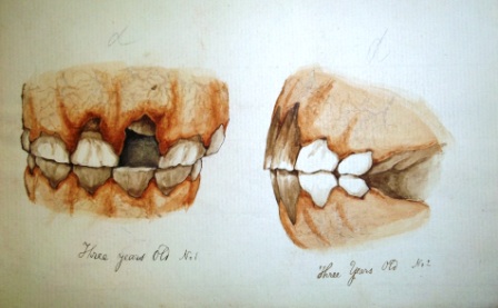
The ones I like best are those drawn by Edward Mayhew – a couple of which are shown below.

The teeth of a three year old horse

Illustration by Edward Mayhew the teeth of a three year old horse

Illustration by Edward Mayhew showing the teeth of a three year old horse

and the  first plate in Mayhew’s book The Horse’s mouth: showing the age by the teeth

Illustration from Edward Mayhew's The Horses mouth: showing the age by the teeth

Illustration from Edward Mayhew’s The Horse’s mouth: showing the age by the teeth 1849

Scabies in the horse – the first Fellowship thesis

I was recently asked the date and subject of our earliest fellowship thesis.  A quick check on the catalogue showed it was written in December 1893 and titled ‘Scabies in the horse: does it demand legislation?

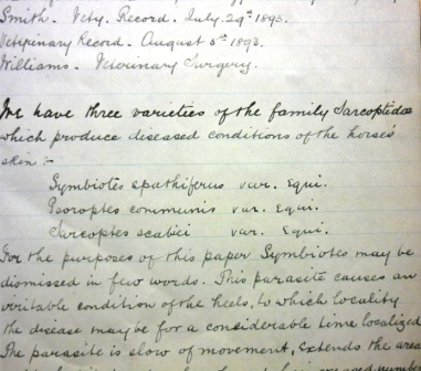
The thesis is a neatly written document Opening linesaccompanied by 12 hand drawn illustrations.  Regulations in place at the time stated that all fellowship submissions be ‘signed by a motto only’ – this one is signed ‘Advance’.

So whose motto was ‘Advance’?  Well the annual report of 1893/94 reveals that the thesis had been submitted by Veterinary Lieutenant R Butler.

Major-General Ernest Reuben Charles Butler (1864-1959) CB CMG DSO FRCVS qualified at the London Veterinary College in 1884 and was gazetted into the Army Veterinary Department in which he served for 37 years. He spent a total of 16 years in India, was an Assistant Professor at the Army Veterinary School, Aldershot 1892-1897 and Professor from 1901-1905.

During World War One he was mentioned six times in dispatches, and made a Companion of the Order of St Michael and St George (1915) and a Commander of the Order of the Bath (1918). In 1919 he was promoted to the rank of Major-General.

Butler also served as an examiner for the RCVS Diploma from 1895 -1904

On retiring from the army at the age of 57 he went to live in Kokstad, KwaZulu-Natal, South Africa where his grave can still be seen today.

The thesis might, at just 16 pages plus bibliography, seem rather short when compared to today’s submissions; in fact it is the standard length for that time.  In it Butler outlines the three varieties of Sarcoptidae which produce diseased conditions of horses’ skin, describes the history of widespread outbreaks of equine scabies and discusses means of accurate diagnosis and the fact that errors may arise from:

‘the eruption being mistaken for a non parasitic form of eczema [or from] the presence of non Psoric Acari on the skin’.

Finally he suggests possible legislative controls that could be put in place.

To aid diagnosis Butler provides illustrations of Acari (see below) drawn from materials he examined.

Filed with the thesis is a letter Butler sent in 1914 asking to borrow it from the library -a request which was granted on the proviso it was returned within a week!

A very common variety

“A very common variety”

Species unknown, found on a healthy horse

“Species unknown, found on a healthy horse”

Debris of Acari

Debris of Acari

Debris of Acari

Debris of Acari

Eclipse and his ‘equine bumps’

In January 1828 the first two British veterinary journals started, these were The Veterinarian and the much shorter lived Farrier and naturalist. Both owed their origins to a desire to comment, and pass critical judgement, on the state of affairs at the London veterinary college. The  Farrier and naturalist is generally believed to be the work of Bracy Clark – it certainly contains a number of unattributed articles that had either previously been, or were later to be, published with Clark as the author.   One of these is ‘A short history of the celebrated racehorse Eclipse’.

Immediately following this is an interesting – again unattributed – article titled ‘Phrenology – it’s utility and importance in animals’ (1)

The article opens with a brief introduction to the ‘science of Phrenology’ and the work of Drs Franz Joseph Gall and Johann Gaspar Spurzheim.   Stating that:

“[Phrenology] is being now approved of and encouraged by men of the highest talents and abilities … we therefore wish to call the attention of our readers to its very great importance … in judging of the character, habits and capacities of different animals.

“A knowledge of Phrenology is of vast importance, as in all cases it will be found that an animal’s courage … sagacity, shyness, meekness, and general temper will depend entirely on the brain; and as this organ is more or less developed, in particular parts, so will the character be found … to correspond to the outward indications of the skull.”

Having set the scene and, hopefully, convinced the reader of the usefulness of Phrenology, the author goes on to describe what Dr Spurzheim found when he examined Eclipse’s skull –apparently Dr S not being a sportsman was not even acquainted with the name of ‘this matchless horse”!

So what did he find?

“A remarkably large brain… strongly indicating great and high courage, unusual sagacity, but deficiency in meekness, or rather a vicious temper”

The author ends by stating that “these peculiarities in his character and disposition will be immediately seen on referring to his history… and the remarkable correctness of the Dr’s observations may serve to show that this science will prove eminently useful in judging of the living animal.”

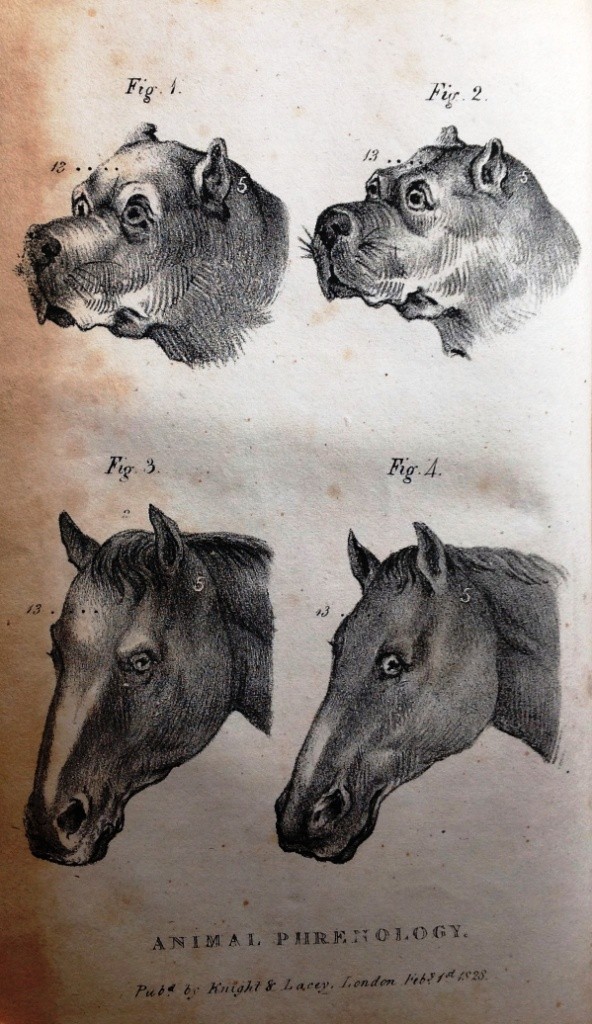
In issue two we find the second part of the article – which opens with this plate

Animal phrenology

Animal phrenology

and explanation

Explanation of plate

Explanation of plate

The numbers refer to Dr Spurzheims classification into organs (there is an explanation of Spurzheim’s organs here) there then follows an explanation of how these can be related to animals.  For example

Secretiveness – propensity, to conceal , or cunning. “where this part of the organ is found large and active in … the Fox….the dog … and the cat, when watching the mouse”

and

Melody – or tune where “the heads and skulls of birds which sing, and of those which do not sing … present conspicuous differences at the place of this organ.”

Convinced?

According to the article in 1828 you could buy a cast of Eclipse’s skull, with the marked organs, from a Mr Deville 367 Strand.   I wonder if any still exist  so we can investigate further?

  1. Farrier and naturalist vol 1 (1) pp34-5, 71-75, 106-109

Strickland Freeman’s observations on the horses foot

Our historical collection has over two hundred books on farriery and horse shoeing many of which contain anatomical drawings of the foot of the horse.  One such book is Strickland Freeman’s

Observations on the mechanism of the horse’s foot its natural spring explained, and a mode of shoeing recommended, by which the foot is defended from external injury, with the least impediment to its spring

which was published in 1796.

Strickland  Freeman was a Buckinghamshire landowner and sporting gentleman  who wrote a number of works on horsemanship and farriery.   Observations is his most important anatomical work: focusing on horse shoeing practices and methods, the work consists of  109 pages of text and 16 coloured plates with outlines.

I have found two reviews of the book from the period.  One in The Critical Review is less than complimentary.   Speaking of the illustrations it says:

“[they] do not illustrate the complete anatomy of the foot:  they are imperfect, from a total neglect of the nerves and absorbent vessels of those parts, two points in the structure, which have been found to be inseparably connected with many diseases of the feet of horses”

The reviewer also takes issue with the cost of the book given its alleged shortcomings:

 “When a voluminous and expensive work is laid before the public…we are led to expect something approaching toward a complete account of that subject… To the anatomist he affords no information, to the gentleman, we fear, he conveys little knowledge which can be applicable in practice… We would advise further adventurers in scientific pursuits with which they are not fully conversant, to be more backward in taxing the world with expensive books”.

A more positive review appeared in The Monthly Review which describes Observations as a ‘most splendid work’ and says:

“it is evidently the result of attentive observation…that will afford useful hints to those who are practically concerned in the subject”.

Writing in 1929 Frederick Smith in Volume 2 of his Early history of veterinary literature says:

“[Freeman] knew nothing of the anatomy of the horse’s foot.  The text is consistently weak, though largely atoned for by…[the] beautifully coloured plates”.

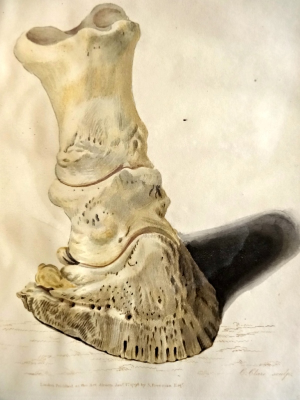
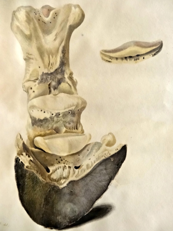
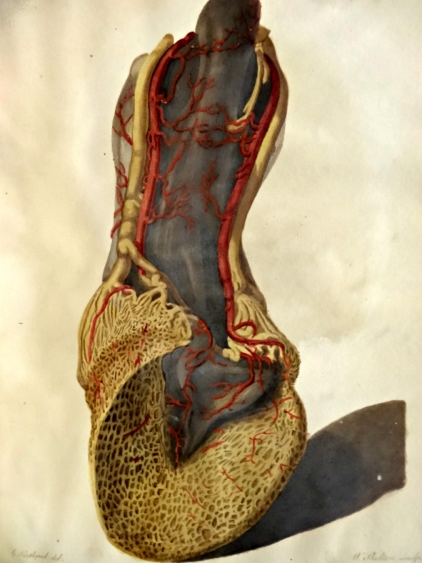
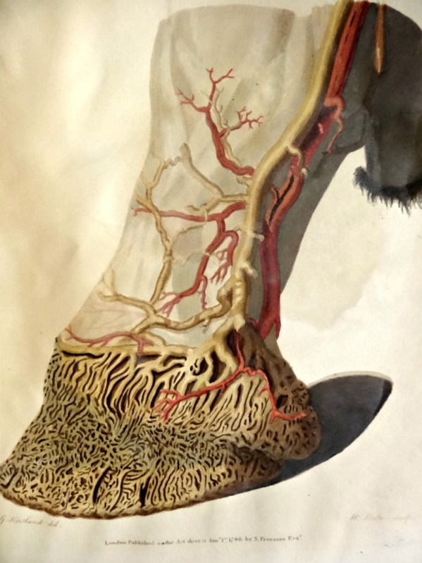
I am not qualified to comment on the accuracy of either the text or illustrations, but can’t help but agree that the illustrations, which are by G Kirtland who was a leading anatomical artist at that time, are beautiful.  Four of them are shown below, what do you think?

Plate 3. Front view of the bones of the fore foot of a horse in their relative situation

Plate 3. Front view of the bones of the fore foot of a horse in their relative situation

Plate 4. Back view of the bones of the fore foot in their relative situation

Plate 4. Back view of the bones of the fore foot in their relative situation

Plate 8. View of the posterior surface of the foot to shew the arteries and veins

Plate 8. View of the posterior surface of the foot to shew the arteries and veins

Plate 9. Side view of the foot to shew the arteries and veins

Plate 9. Side view of the foot to shew the arteries and veins

Shining a light on veterinary artists

The Lightbox in Woking, is currently showing an exhibition describing the role of horse and mules in World War 1.  The Horse at War: 1914-18  has a wide ranging display of artwork both from the war itself as well as more recent works, most noticeably ‘Joey’ the life size puppet from the National Theatre’s stage production of War Horse. Amongst the many paintings on display are works by a number of official war artists including Sir Alfred Munnings, CRW Nevinson and Lucy Kemp Welch.

There are a number of interesting veterinary connections amongst the paintings.  We have loaned two paintings by Lionel Edwards .  Born in Bristol in 1878, Edwards was one of the most popular illustrators of hunting and sporting subjects of the twentieth century. His artistic talents were apparent early in life, drawing horses from the age of six. He studied in London at Heatherly’s School of Art, before pursuing a professional career as an artist. At the outbreak of war he enlisted and served as a Remount Purchasing Officer, responsible for purchasing horses, an experience he described as ’four solid years of nothing but horse.’

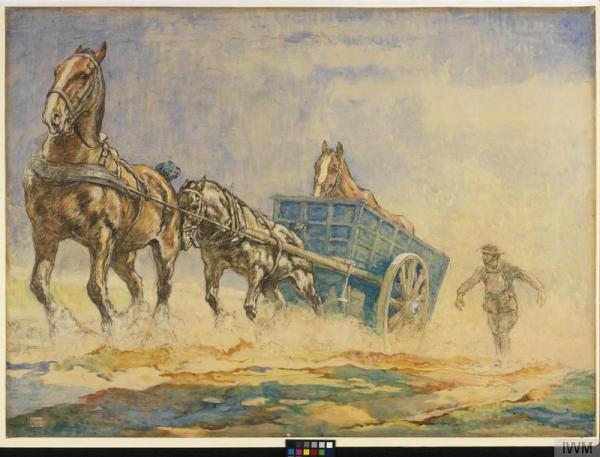
Also on display are a number of works by official war artist Edwin Noble.  Noble studied at the Slade School of Art and the Royal Academy and became an established illustrator prior to the war.  He served with the Army Veterinary Corps, rising to the rank of sergeant, spending much of the war at No 8 Veterinary Hospital in France. Here he recorded in great detail the diseases affecting horses, ranging from mange through to the effects of mustard gas. His pictures were described as providing an ‘almost veterinary eye view of the misfortunes of horses on active service.’

A Horse Ambulance Pulling a Sick Horse out of a Field by Edwin Noble

A Horse Ambulance Pulling a Sick Horse out of a Field by Edwin Noble © IWM (Art.IWM ART 2922)

There are three works of art on display produced by a veterinary surgeon, but one who was serving with the Royal Army Medical Corps during the conflict.

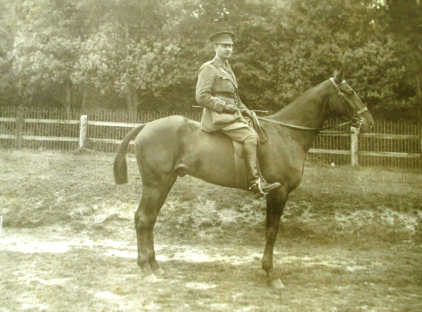
Herbert Lake was born in 1883, at 33 High Street Camden, London, where his father owned a jewellers shop. Qualifying from the Royal Veterinary College in 1905, he initially worked in London passing the examinations for an Inspector of Meat and other Foods, in December 1908. Soon after he entered University College London to read medicine, gaining MRCS, LRCP in 1913 and MB in 1915. On graduation from UCL Lake joined the Royal Army Medical Corps and was posted to northern France in October 1915, to serve as medical officer to 2 Cavalry Field Ambulance (2CFA) (part of 2nd Cavalry Division). War diaries show how he used both his medical and veterinary knowledge.

Herbert Lake 1883-1869

Herbert Lake 1883-1969

Soon after his arrival, responding to the cold and wet conditions, Lake noted that horses had been standing out all the time and though there had been no sickness among them, they had generally fallen off in condition. To provide protection he moved horses into the trenches. It was recorded that ‘it has not been entirely successful owing to the heavy mud, but they have been sheltered in this way from the cold winds.’ Two months later his veterinary skills were again to the fore, when he was asked to give a course of lectures in horse mastership and stable management to each field ambulance.

However it was his medical work that was to be recognised when on 8 October 1916 he led a digging party in the Ginchy area of the Somme. The party dug out three men; one was dead, the other two wounded. Lake would later be mentioned in dispatches for his actions.

Herbert Lake was also an accomplished artist, although unlike many of his contemporaries, he appears not to have had any formal training.  Making use of a number of media, he portrayed his experiences in war, and invariably horses are at the centre of his work. He graphically depicted the role of the field ambulance in a number of sketches.

Horse pulled ambulance by Herbert Lake 1917

Horse pulled ambulance by Herbert Lake 1917

In March 1917, Lake witnessed the cavalry charge at Arras, the last cavalry charge by British troops in Europe. His painting ‘Cavalry Before Arras’ portrays the intensity of the preparations for battle amongst both horse and rider. Later Lake would treat many of those injured in the battle,

Cavalry before Arras by Herbert Lake

Cavalry before Arras by Herbert Lake

After the war Herbert Lake settled in Beaminster, Dorset where he established a general medical practice. However, he remained on the register of the Royal College of Veterinary Surgeons until 1958, and treated patients, both human and animal, for many years.

The exhibition runs until 1 March.

Acknowledgements
I would like to thank Dr Paul Watkins MRCVS for his help in compiling this post.

Images

A Horse Ambulance Pulling a Sick Horse out of a Field by Edwin Noble © IWM (Art.IWM ART 2922)   reproduced under the IWM Non-commercial Licence

Photograph of Herbert Lake and the paintings by him are reproduced with the permission of the Lake family.

A Gift Horse

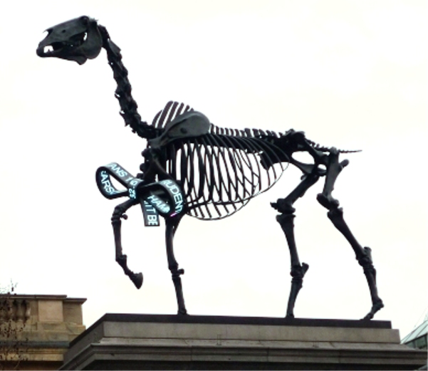
Gift Horse by Hans Haacke, Trafalgar Square

Gift Horse by Hans Haacke, Trafalgar Square

Sitting proudly on top of the fourth plinth in Trafalgar Square currently is ‘Gift Horse’ by Hans Haacke.

The sculpture is based on illustrations from George Stubb’s Anatomy of the horse and so feels strangely familiar to me – apart from the live share-price ticker tied to its foreleg.

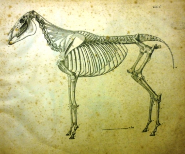
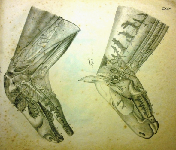
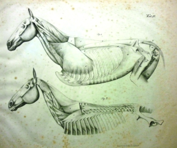
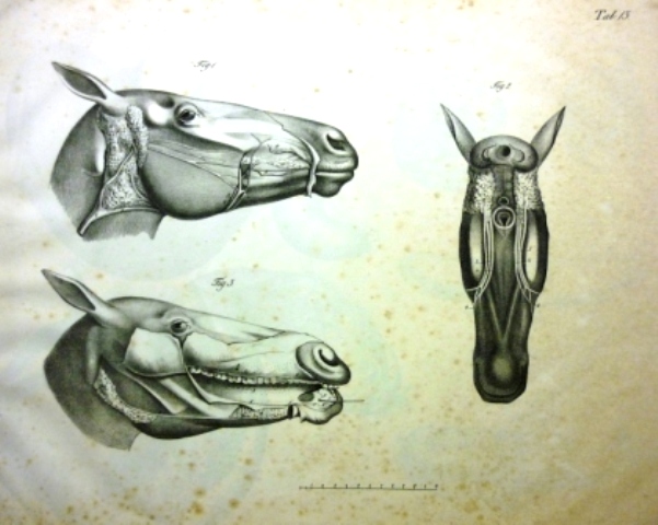
Looking at our catalogue I found we have another book of the same title in our collection – Ernst Gurlt’s  Anatomy of the Horse (A Schloss 1833, translated from the original German by J Willimott) and like the Stubbs it is lavishly illustrated.

Ernst Friedrich Gurlt (1794-1882)  was Professor at the veterinary school in Berlin where the anatomy collection  still bears his name.

He published a number of works on anatomy the first of which was Handbuch der vergleichenden Anatomie der Haus-saugethiere  (Handbook of comparative anatomy of domesticated animals).  This covered the anatomy of the horse, ox, sheep, swine, dog and cat.

A review of the English translation of Gurlt’s Anatomy of the Horse in the Veterinarian (1833 pp 46-50) begins by setting Gurlt’s anatomical works in the context of those that had gone before describing  his  Handbuch der vergleichenden Anatomie der Haus-saugethiere ‘as the great treasure of the veterinary anatomist’.

Of the English translation of the Anatomy of the Horse it is less favourable – though no discredit is attached to Gurlt.  Instead it argues that by taking the plates on the horse from Gurlts more comprehensive earlier book of comparative anatomy  the publisher, Mr Schloss, has ‘destroyed the charm of the work, as one of comparative anatomy and rendered it incomplete as an elementary book’.

The review does go on to say however that ‘to the student who confines his inquiries to the horse, it is still a most valuable present’.   The cost of this ‘valuable present’ is given as £3 5s.

Anatomy of the horse was published in two parts – the text which is an index to the plates (128 p 8vo) and the 35 folio sized plates several of which are reproduced below.

Gurlt Plate 1 Skeleton seen from the left sidePlate 1 Skeleton seen from the left side

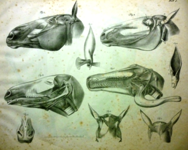
Gurlt Plate 7 Muscles of the headPlate 7 Muscles of the head

Gurlt Plate 10 Fourth layer of muscles seen from the left sidePlate 10 Fourth layer of muscles seen from the left side

Gurlt Plate 13 Salivary glandsPlate 13 Salivary glands

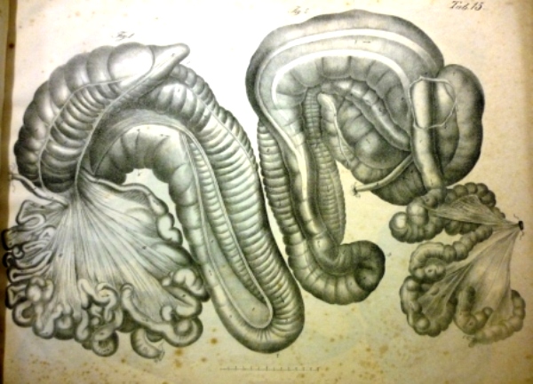
Gurlt Plate 15 Intestinal canalPlate 15 Intestinal canal

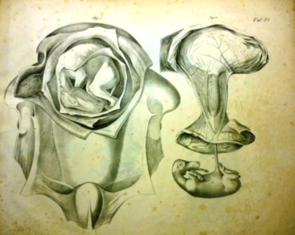
Gurlt Plate 22 Pregnant uterus of the mare, opened from belowPlate 22 Pregnant uterus of the mare, opened from below

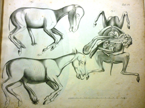
Gurlt Plate 24 Female foetus 57 days oldPlate 24 Female foetus 57 days old

Gurlt Plate 28 arteries on the head and neck

Plate 28 Arteries on the head and neck