The many layers of the dog
There has been a lot of discussion of anatomical illustrations recently following the opening of the exhibition at the Queen’s Gallery Leonardo da Vinci anatomist. We have a number of stunning anatomical illustrations in the Historical Collection – the most well known would be in Stubbs’ The anatomy of the horse and then there are those in Carlo Ruini’s Anatomia del cavallo, infermita, et suoi rimedii and Andrew Snape’s Anatomy of an horse.
Although I admire these works my favourite anatomical items are the books of models that we have. Turn the page and lift the flaps to see what lies behind – it is like being a child again.
We have two complete series of these ‘flap books’: Vinton’s livestock models – which includes the pig, the sheep, the bull, the cow, the horse, and the mare and foal and Philips’ anatomical and technical models, series 2.
This second set by Philips covers domestic animals. Again we find the horse, the ox, the sheep and the pig but interestingly we also find the dog. Why interestingly? Well these books were produced in the 1890s, a time when the practice of keeping a dog as a companion animal was not common.
The dog: its external and internal organisation: an illustrated representation and brief description was edited by Alexander Constant Piesse MRCVS who graduated from the Royal Veterinary College in 1885 and the anatomical description is by William S Furneaux.
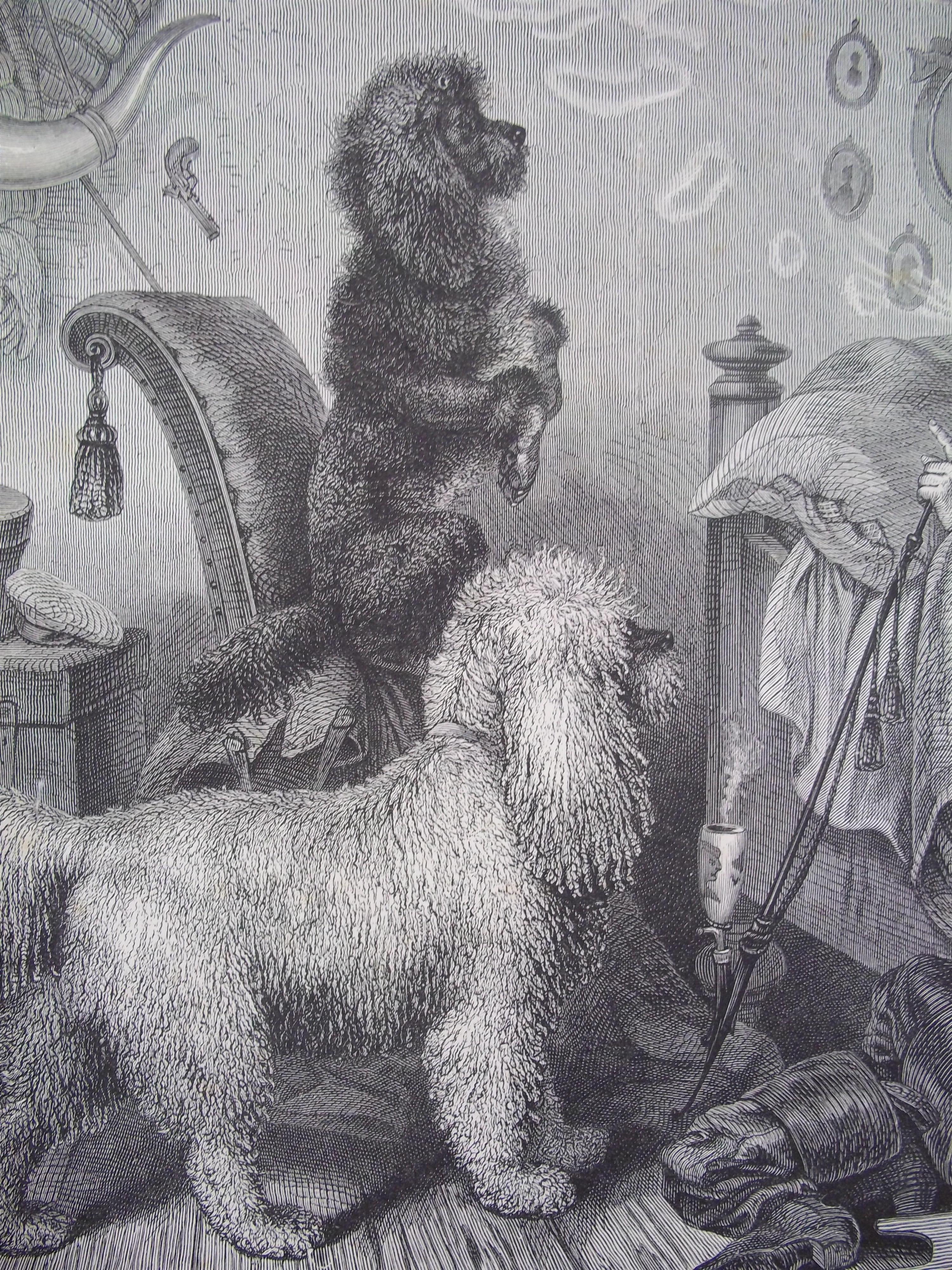
It starts with a 28 page text which gives the general characteristics of dogs, then divides dogs into two types – non-sporting and sporting and gives the characteristics of the main breeds. This section is illustrated with some rather nice images.
There then follows “a detailed account of the anatomy of the body of the dog; [which] will be illustrated by means of a folding model of the St Bernard”. The various ‘layers’ are then discussed – starting with the exterior, the skin, and ending with the genital organs.
Finally, there is an explanation of the folding plates with each section numbered to correspond to the model. So we have: Head – (1) Nose (2) Crest of nose (3) Mouth and upper lip etc
There are 5 folding plates in all – and you lift the flap to reveal the next ‘layer’. The inner ‘layers’ are a mass of flaps – so much so that instructions are given as to how to open them “No 13 [left lung] may be turned upward and 18 [left ventricle] to the left …the interior of the hear them become[s] visible”.
Please get in touch if you want to visit and explore the layers of the dog – or horse or pig ….



Leave a Reply
Want to join the discussion?Feel free to contribute!